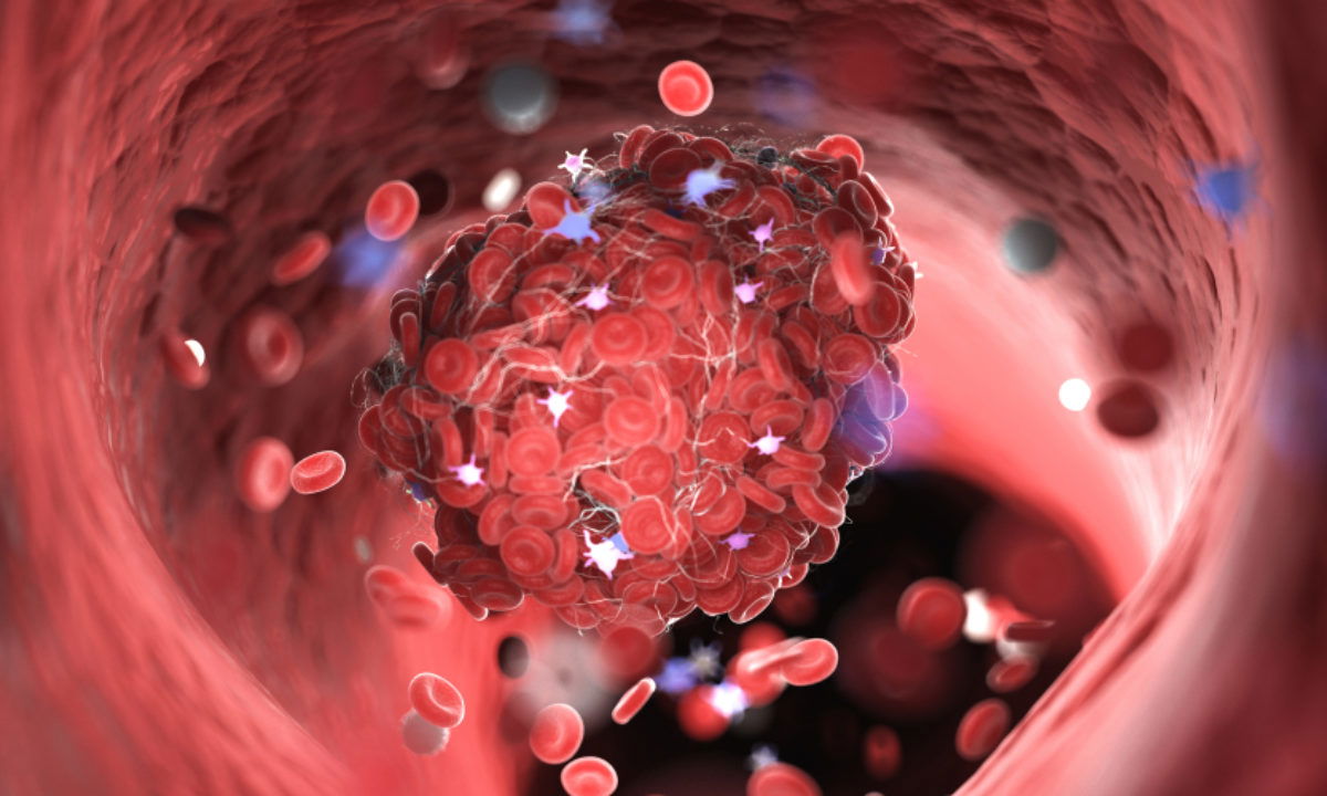
وفاه مريضه بعد التحسن من الكوڤيد ١٩ بسبب قلبي نتيجه المتلازمة الاتهابيه متعددة الأجهزة

في بعض المرضى ، يمكن أن يؤدي الاصابه بالكوفيد 19 الى تشكل جلطات دموية مجهرية في الشعيرات الدموية والشرايين الصغيرة مما قد يؤدي إلى اذيه او تلف في بعض الأعضاء ، أو قد يؤدي الى متلازمة التهابية متعددة الأنظمة تشبه مرض كاواساكي. يتضمن التقرير الحالي مريضه أصيبت بـ COVID-19 وتعافت ولكنها رجعت الى المشفى مره ثانيه نتيجه التهاب في الأوعية الدموية الصغيرة للقلب أدت الى الوفات بشكل سريع في قسم الطوارئ. نشرت الحاله في مجله Annals of Internal Medicine.
كانت المريضة امرأة من اصل أفريقي وتبلغ من العمر 31 عامًا ولديها قصه من السمنة وارتفاع ضغط الدم والسكري ( غير المضبوط، مخزون السكر ١٤٪). جاأت الى غرفه الطوارئ تشكو من الحمى والسعال الجاف وألم في البطن لمدة 5 أيام. ثبتت إصابة المريضة بـ SARS-Cov2 وتم قبولها الى المشفى بتشخيص COVID-19. عولجت بأزيثروميسين ويومين من هيدروكسي كلوروكين وبعدها شعرت بالتحسن وتم تخريجها من المشفى بعد ان زالت الحمى وأصبحت نسبه اوكسجين الدم 95٪ وهي تتنفس هواء الغرفة.
عادت المريضه بعد 12 يومًا بحمى مفاجئة، و آلام نابضة في الجانب الأيسر من الرقبه مع الغثيان والقيء. كانت تعاني من الحمى التي بلغت 39.8 درجة مئوية ، وكان لديها تسرع قلب جيبي بمقدار 120 نبضة / دقيقة.
اظهر الفحص البدني التهاب وتورم في الغدة النكفية. أظهر الفحص بالتصوير الطبقي المحوري لرقبتها تضخمًا ثنائيًا في الغدد النكفية وتورم في البلعوم الأنفي الخلفي و الفموي ، وأظهر الفحص بالتصوير الطبقي لصدرها تحسنًا في آفات الرئة الارتشاحيه ، ولكن مع تضخم في العقد الليمفاوية العنقية والأمامية للمنصف. كان فحص PCR لمسحة البلعوم سلبية لـ SARS-CoV-2. أظهرت النتائج المخبرية ارتفاع عدد خلايا الكريات البيضاء ومستوى D-dimer ٢،٤٨ نانومول / لتر و كان هناك ارتفاع في CRP. أثناء تقييم المريضه في غرفه الطوارئ لقبولها الى المستشفى ، أصيبت بعدم استقرار الدورة الدموية hemodynamic instability والرجفان البطيني ولم يتمكن الاطباء من إنعاشها. تم تشريح الجثة في محاولة لتحديد السبب الرئيسي للوفاة. تضمنت النتائج التي توصل إليها تشريح الجثة تضخم العقد الليمفاوية في العنق والمنصف ، وتجلط الدم الوعائي مع نزيف محوري محيط في الرئة اليسرى السفلية ، والتي ربما ساهمت في المرض ولكن لم يكن على الأغلب السبب الرئيسي للوفاة. أظهر الفحص المجهري للرئة وجود نزيف حاد بؤري. تم العثور علىاظهر الفحص المجهري للغدد الليمفاوية العنقية تغيرات تفاعلية reactive فقط. كان مظهر القلب طبيعي، وبدون دليل على توسع في الشرايين الاكليليه أو تصلب أو تضيق في الشرايين. ولكن مجهرياً كان هناك التهاب في بطانة الأوعية الدموية والتهاب في الأوعية بشكل منتشر في الأوعية القلبية الصغيرة ويمتد إلى الشحم المحيط بالقلب والتامور (الشكل ،A ،B). لم يكن هناك ارتشاح للخلايا الليمفاوية في عضلة القلب. التهاب الأوعية الدموية تظاهر بوجود العديد من الكريات المعتدلة neutrophils (الشكل ، C) ، وكذلك وجود خلايا CD4 > CD8 الليمفاوية (الشكل ، E و F). لم يكن الالتهاب موجودًا في الشرايين التاجية أو الأوعية الدموية الكبيرة (الشكل ، D). لوحظ التهاب مماثل في أوعية الكبد (الشكل G)
تظهر هذه الحاله استجابه المريضة للعلاج الأولي في المستشفى ، إلا أنها تعرضت لاحقًا لعقابيل شملت القلب (والأعضاء الأخرى). وتدهور سريع نتيجه تضرر في القلب بعد أسبوعين تقريبًا من خروجها من المستشفى.
الصورة السريرية لاعتلال العقد اللمفية المفاجئ والتهاب الغدة النكفية المقترن بالتهاب الأوعية القلبية الصغيرة بعد COVID-19 توحي بشدة بمتلازمة التهابية متعددة الأنظمة multisystem inflammatory syndrome. كان سبب الوفاة هو التهاب الأوعية الدموية ، الذي يُفترض أنه ادى الى اضطرابات النظم القلبيه القاتلة ، ولم يكن هناك دليل على التهاب عضلة القلب.
يسلط التقرير الضوء على احتمالية حدوث مضاعفات خطيرة بسبب تلف بطانة الأوعية الدموية مما يؤدي إلى متلازمة الالتهاب متعددة الأجهزة بعد COVID-19 ، وهو مشابه محتمل لالتهاب عضلة القلب الحقيقي. لذلك يجب المراقبة الدقيقة للعلامات المختبرية للالتهاب في مرضى الكوڤيد ١٩، وكذلك يجب التدخل العلاجي مثل إعطاء الكورتيزون، لمعالجه هذه العملية الالتهابية ، فلربما تؤدي إلى تحسين نتيجه الم
https://www.acpjournals.org/doi/10.7326/L20-0882
Death of an adult woman after recovering from covid-19 due to cardiac etiology as consequence of the development of multisystem inflammatory syndrome
In some patients, covid 19 can be associated with microscopic blood clots in capillaries and small arteries leading to organ damage, or with a multisystem inflammatory syndrome that is similar to Kawasaki disease. The current report involves a patient with COVID-19 and vasculitis of the small vessels of the heart who died in the emergency department suddenly.
The patient was a 31-year-old woman with history of obesity, hypertension and diabetes (poorly controlled, Hb A1c 14%). She was admitted for fever, dry cough, and abdominal discomfort of 5 days. Patient tested positive for SARS-Cov2 and was admitted with COVID-19. She was treated with azithromycin and 2 days of hydroxychloroquine and at discharge, she was afebrile and her oxygen saturation was 95% on room air.
The patient returned 12 days later with sudden fever; throbbing, left-sided neck pain; nausea; and vomiting. She had a fever of 39.8 °C, with sinus tachycardia of 120 beats/min.
Her physical examination was remarkable for parotitis. A CT scan of her neck showed bilaterally enlarged parotid glands and swelling in the posterior nasopharynx to oropharynx, and a CT scan of her chest showed improvement of her lung lesions, but with cervical and anterior mediastinal lymphadenopathy. PCR of nasopharyngeal swab was negative for SARS-CoV-2. Laboratory results showed an elevated white blood cell count , a D-dimer level of 2.48 nmol/L, and elevated CRP. While she was being evaluated for hospital admission, she developed hemodynamic instability and ventricular fibrillation and could not be resuscitated. An autopsy was done in an attempt to determine the primary cause of death. Finding on autopsy included enlarged cervical and mediastinal lymph nodes, and vascular thrombi with focal surrounding hemorrhage in the left lower lung, which probably contributed to illness but were not likely the primary cause of death. Pulmonary microscopic examination showed focal acute hemorrhage and numerous megakaryocytes. Cervical lymph nodes were found to have reactive changes. The heart had a grossly normal appearance, without evidence of coronary artery aneurysm, atherosclerosis, or stenosis. Microscopically, however, endotheliitis and vasculitis were present, diffusely involving the small cardiac vessels and extending into the surrounding epicardial fat and interstitial spaces (Figure, A and 😎. There was no lymphocytic infiltrate of the myocardium. The vasculitis was composed of numerous neutrophils (Figure, C), as well as CD4+>CD8+lymphocytes (Figure, E and F). Inflammation was not present in the coronary arteries or larger blood vessels (Figure, D). Similar inflammation was noted in occasional portal triad vessels within the liver (Figure, G). This patient survived an initial hospitalization, however she had later sequelae involving the heart (and other organs). the patient had a rapid decompensation and evidenced heart damage almost 2 weeks after discharge from her initial hospitalization. The clinical picture in of sudden lymphadenopathy and parotitis combined with small-vessel cardiac vasculitis after COVID-19 is strongly suggestive of a multisystem inflammatory syndrome. The cause of death was a vasculitis, presumably inciting a fatal arrhythmia, without evidence of myocarditis. The report highlights the potential for serious complications due to endothelial damage leading to multisystem inflammatory syndrome after COVID-19, a possible mimicker of true myocarditis. Careful monitoring of laboratory markers of inflammation, as well as therapeutic intervention such as steroids to target this inflammatory process, may improve patient outcomes. (Annals of Internal Medicine 2020)
د.سوسن جلعوط